MedOne Education
Anatomy Titles
Stunning anatomy atlases loved by students!
Select Thieme Anatomy titles are available via MedOne Education, Thieme’s medical learning platform comprised of medical textbooks as a part of it’s premium titles package. These titles can be licensed on a title-by-title basis or together as a package.
Why Thieme Anatomy?
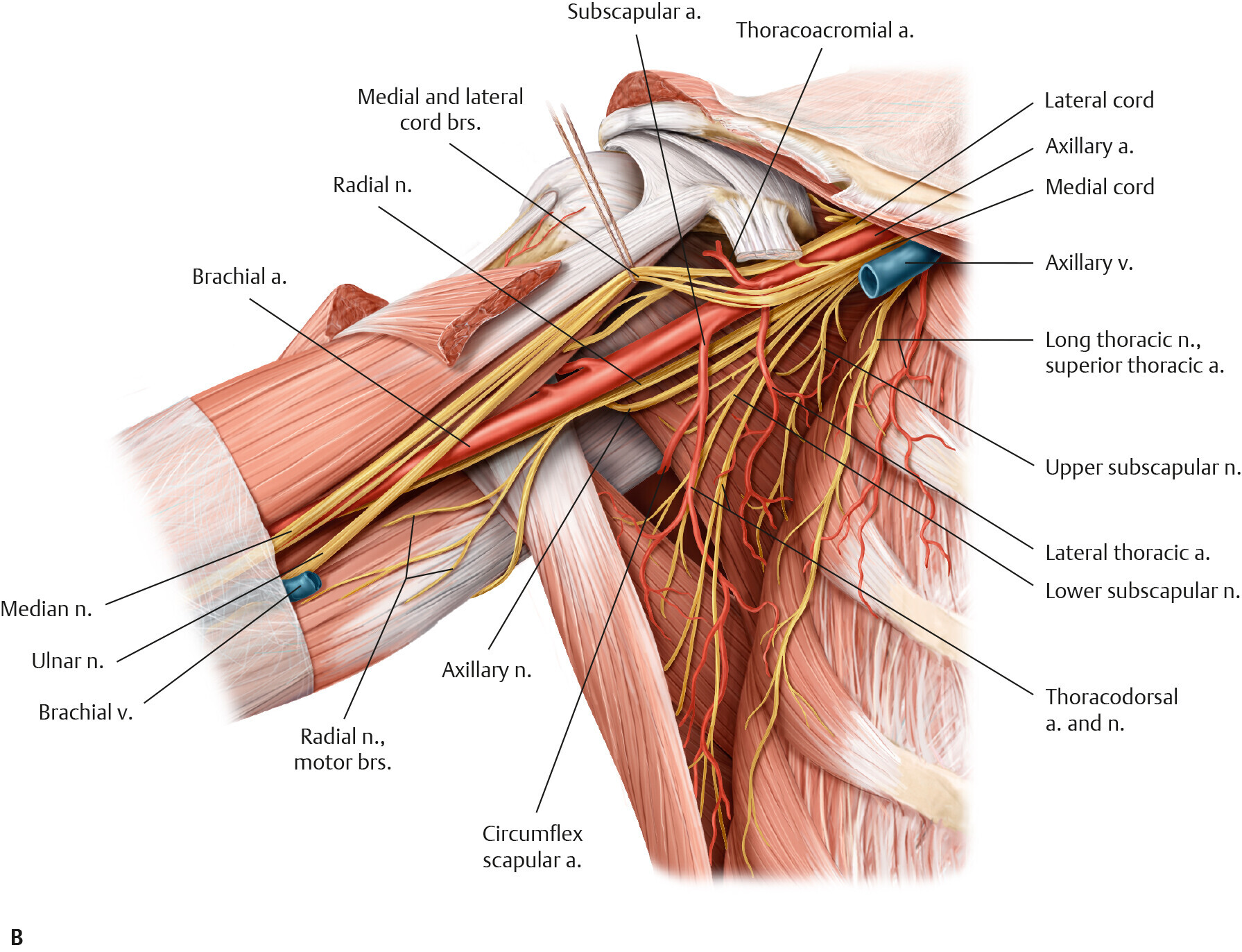
Thieme’s Anatomy titles contain everything students need to successfully tackle the daunting challenges of anatomy. Covering topics such as Basic Anatomy, Dental Medicine, General Anatomy and the Musculoskeletal System, Neck and Internal Organs, and Head and Neuroanatomy, students can hone in on specific topics and focus on their chosen specialties. All titles integrate stunning, full-color anatomy illustrations with must-know clinical concepts on every page, along with expertly rendered cross-sections, x-rays, and CT and MRI scans to vividly demonstrate clinical anatomy.
This visually stunning set of atlases is an essential companion for medical students or residents interested in an in-depth study of anatomy and neuroanatomy for laboratory dissection and clinical reference. This collection is a must-have for allied health students, instructors, and practicing physical and massage therapists, it also serves as a wonderful anatomic reference for professional artists and illustrators.
Available Anatomy Titles
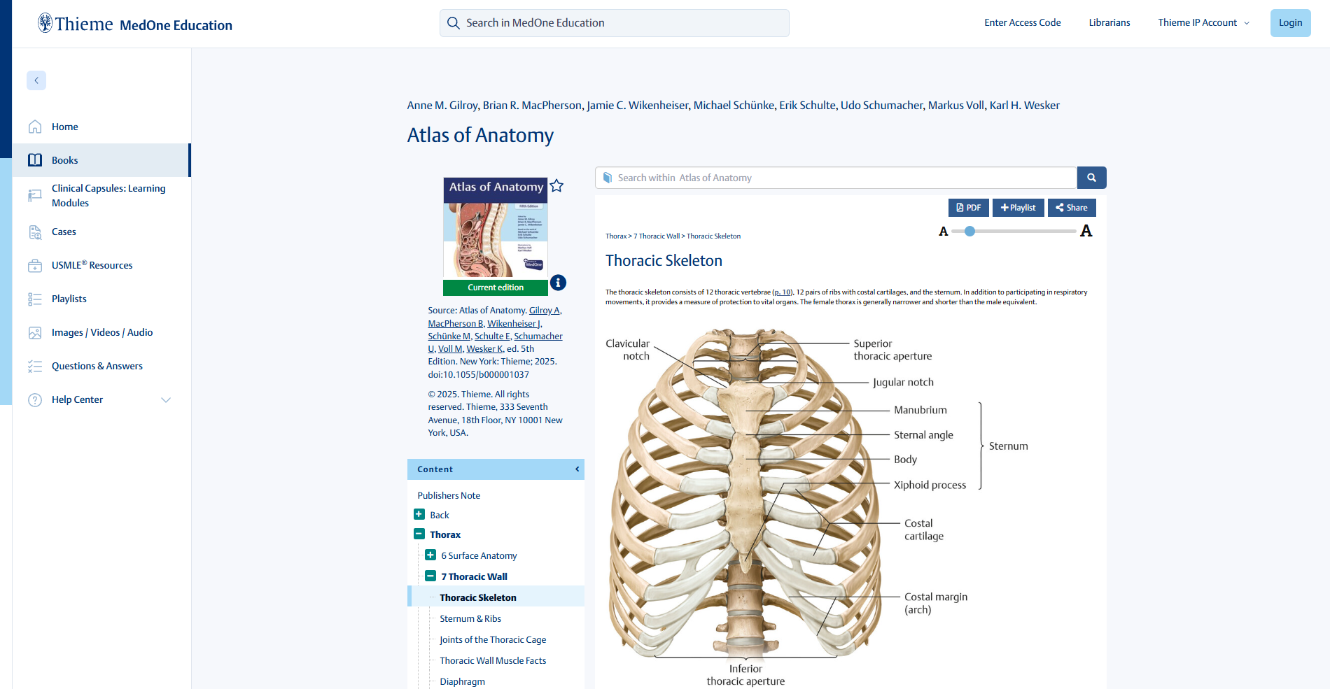
Atlas of Anatomy
Gilroy | MacPherson | Wikenheiser | Schuenke | Schulte | Schumacher
Publication Date: May 2025 | Edition: 5 | 778 pp, 2,113 illustrations
Print ISBN: 9781684206650 | E-Book ISBN: 9781684206681
The definitive resource for learning and teaching challenging anatomy
Rapid advances in medical research and technology have led to innovative medical procedures, and in turn, emerging trends in medical education. Novel developments highlight the importance of once seemingly irrelevant anatomic details, while also impacting the scope of anatomy curricula. To reflect these changes, each chapter in Atlas of Anatomy, Fifth Edition has been meticulously updated to ensure content is both relevant and reflective of the role of anatomy in contemporary medical practice.
The book features seven anatomical sections with a total of 51 chapters, with updates, additions, and revisions incorporated throughout the more than 700 pages. The Table of Contents is enhanced with reader-friendly chapter and page references for all tables and clinical boxes. To mirror the current trend in muscular attachment terminology, origin and insertion are replaced with the more accurate and descriptive terms superior attachment/inferior attachment or proximal attachment/distal attachment. Over 2,100 illustrations unmatched in both the realistic depiction of anatomy and the three-dimensionality of the figures enhance recognition and memorization of structures.
Key Features:
- Reorganization of the neurovascular spreads in the abdomen and pelvis units
- Expansion of the sections on the female breast and perineum
- Added brainstem cross section layouts
- A list of common eponyms with their official anatomic equivalents
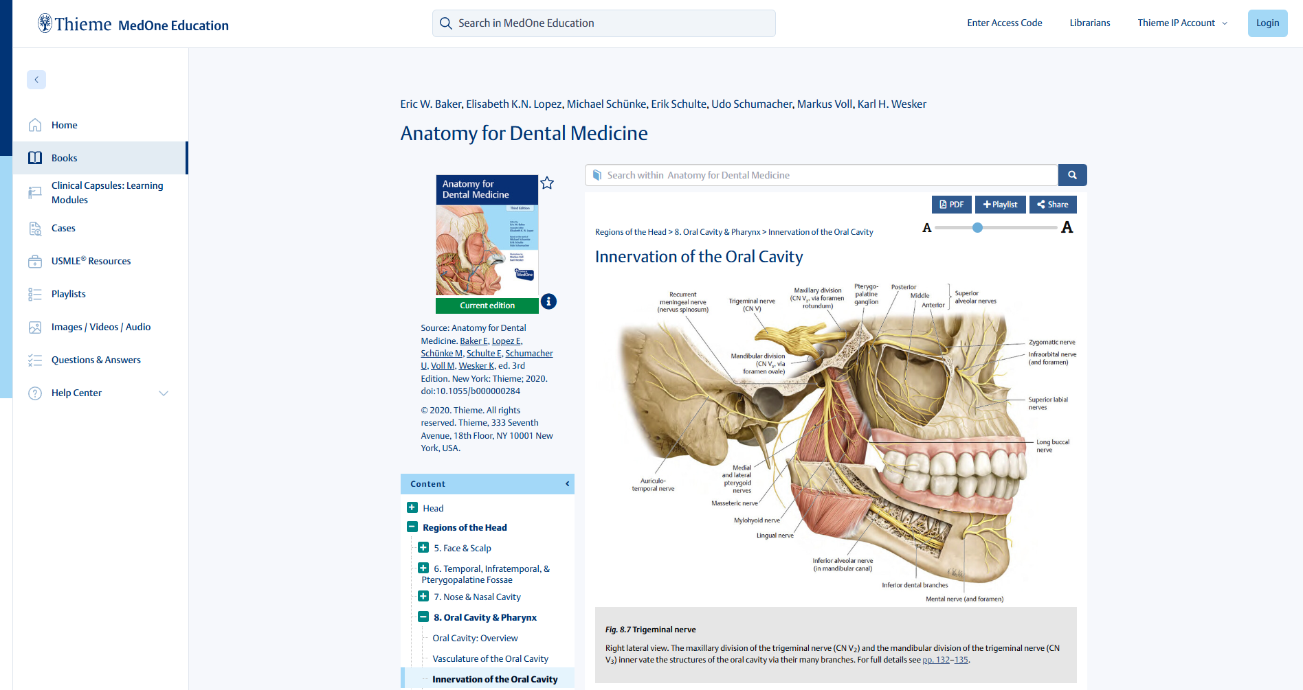
Anatomy for Dental Medicine
Schuenke | Schulte | Schumacher | Baker
Publication Date: July 2020 | Edition: 3 | 608pp, 1,262 illustrations
Print ISBN: 9781684200467 | E-Book ISBN: 9781684200474
Anatomy for Dental Medicine, Third Edition strikes an optimal balance between systemic and regional approaches to complex head and neck anatomy. Award-winning full-color illustrations, succinct text, summary tables, and questions put anatomical structures and knowledge into a practical context.
This is an essential resource for dental students and residents that also provides a robust review for the boards or dissection courses.
Key Features:
- Additional radiologic images and landmark features throughout
- Reorganized brain/nerve sections
- Expanded clinical question appendix including patient box questions in the style of the INBDE
- Factual question appendix places greater emphasis on areas including the skull, larynx, cross sectional anatomy, body below the neck, and local anesthesia
- 1,200 clear and detailed full-color illustrations
- 150 tables for rapid access to key information
- Expanded captions detail key information and clinical correlations
- Appendix covering anatomy for local anesthesia
- Online images with “labels-on and labels-off” capability are ideal for review and self-testing
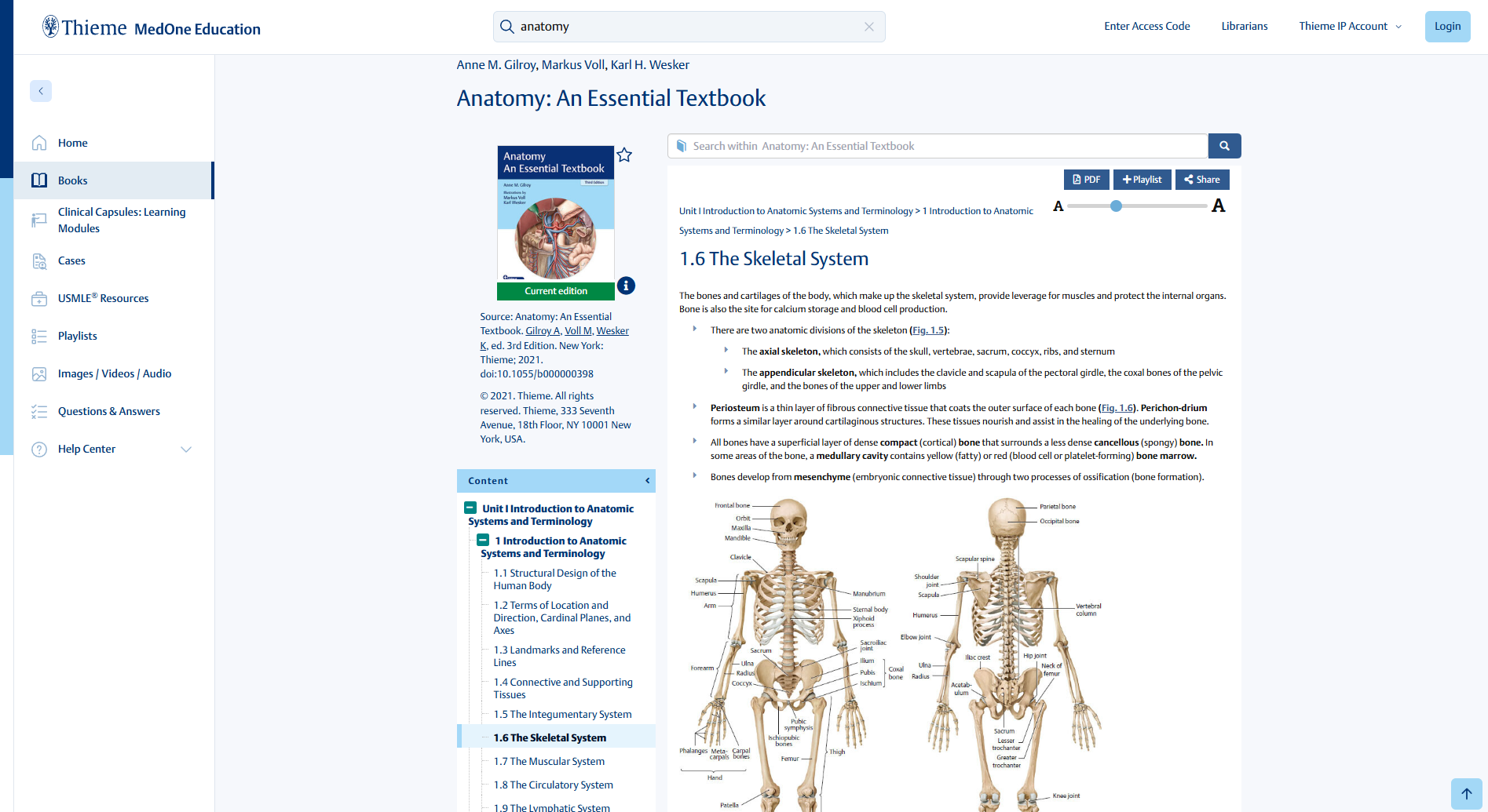
Anatomy: An Essential Textbook
Gilroy | Voll | Wesker
Publication Date: July 2021 | Edition: 3 | 640 pp, 735 illustrations
Print ISBN: 9781684202591 | E-Book ISBN: 9781684202607
Organized by eight units, the book starts with basic concepts and a general overview of anatomic systems. Subsequent units focused on regional anatomy cover the Back, Thorax, Abdominal Wall and Inguinal Region, Pelvis and Perineum, Upper Limb, Lower Limb, and Head and Neck. Each unit includes a chapter on the practical application of regional imaging and extensive question sets with detailed explanations. A new ordering of chapters now mirrors the revised organization of the Atlas and sequence of dissections in most gross anatomy programs.
Key Features:
- More than 100 new images, updated illustrations, and revised versions of all autonomic schematics enhance understanding of anatomy
- New topics in clinical and developmental anatomy addressed throughout include clinically important vascular anastomoses, spinal cord development, and common anatomic anomalies
- Matching colored side tabs allow quick access to similar units in both books
- Over 50 of the new and previously included clinical and developmental correlations now feature descriptive images, radiographs, or schematics
- Self-testing sections in each unit have been expanded with over 40 new USMLE-style question sets with detailed explanations
This is the quintessential resource for medical students to build anatomy knowledge and confidence as they progress in their medical careers.
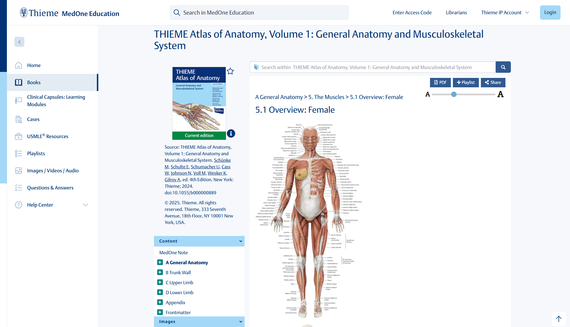
THIEME Atlas of Anatomy, Vol. 1
General Anatomy and Musculoskeletal System, 4th Edition
Schuenke | Schulte | Schumacher | Johnson
Publication Date: 2025 | Edition: 4 | 658 pages, 2120 illustrations
Print ISBN: 9781684205899 | E-Book ISBN: 9781684205998
An exceptional, beautifully illustrated resource on general anatomy and the musculoskeletal system
The unique atlas is divided into four major sections, starting with General Anatomy, which lays a fundamental groundwork of knowledge—from human phylogeny and ontogeny to general neuroanatomy. The three subsequent sections, the Trunk Wall, Upper Limb, and Lower Limb, are systemically organized, presenting bones, ligaments, and joints; musculature; and neurovascular, followed by topographical overviews in each group. Anatomic concepts and clinical applications are introduced in a step-by-step sequence through illustrations, succinct explanatory text, and summary tables, thereby supporting classroom learning and active dissection in the laboratory.
Key Features:
- Female skeletal muscles, genital structures, and surgical interventions, with a new section on muscle fasciae
- More than 2,100 extraordinarily accurate and beautiful illustrations by Markus Voll and Karl Wesker, including a significant number revised to reflect gender and ethnic diversity
- Clinically important musculoskeletal anatomy and pathology imaging for plain film, CT, and MRI scans
- A new chapter on muscle fasciae structure and function covers innervation, compartment syndrome in the lower leg, and classification of the fasciae of the trunk and body cavities
- Variants in human anatomy, such as blood vessels whose courses deviate from the “norm,” or anomalous positions of organs
The updated edition of this best-selling atlas is an essential tool for physical therapy and osteopathic medical students and instructors. It is also an outstanding reference for chiropractors, practicing physical and massage therapists, yoga instructors, and professional artists and illustrators.
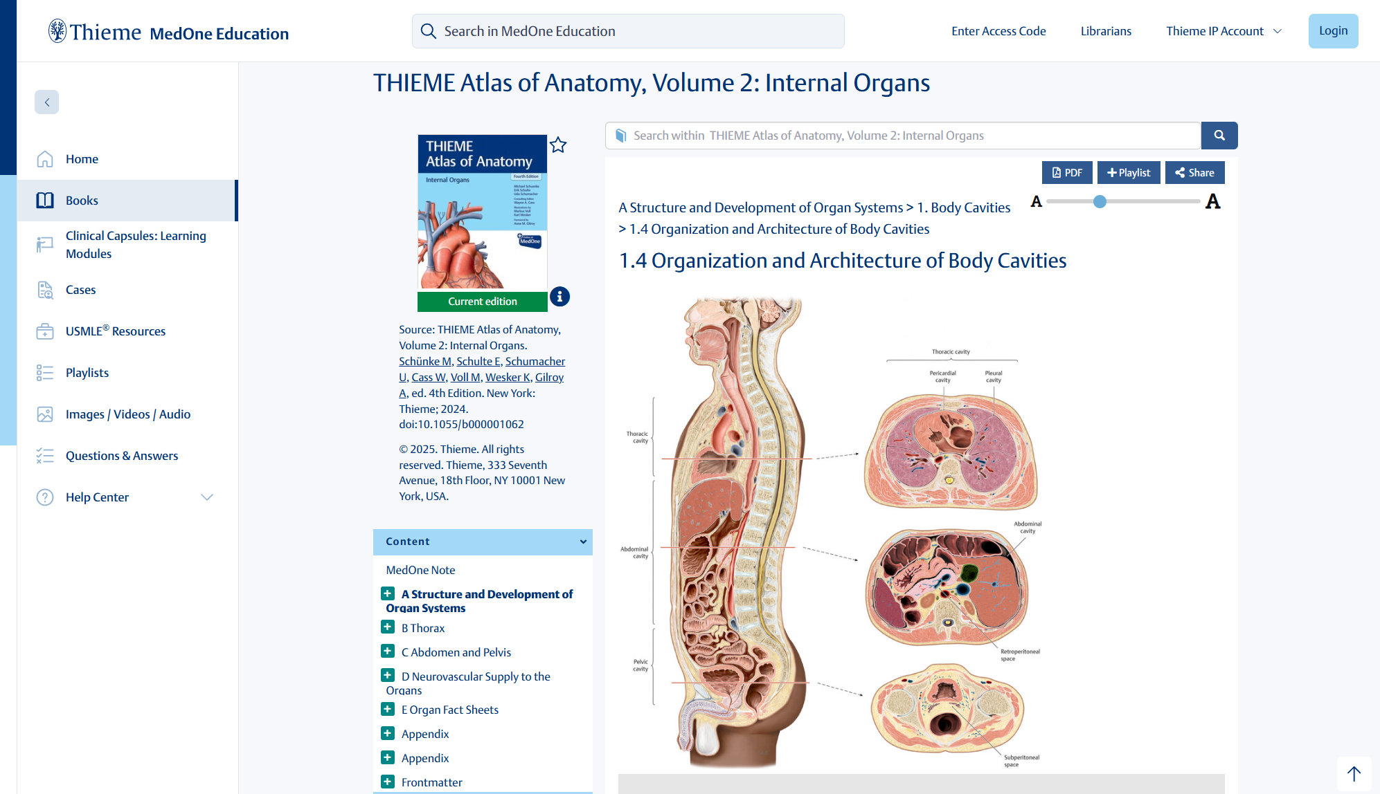
THIEME Atlas of Anatomy, Vol. 2
Internal Organs, 4th Edition
Schuenke | Schulte | Schumacher | Cass
Publication Date: 2025 | Edition: 4 | 538pp, 1,437 illustrations
Print ISBN: 9781684205912 | E-Book ISBN: 9781684205936
Extraordinarily detailed, step-by-step anatomic atlas informs learning and medical practice
The book lays a solid foundation of anatomic knowledge, with an introductory overview of each of the organs, including discussion of embryologic development. Each of the two regional units starts with a short overview chapter, followed by the structure and neurovasculature of the region and its organs. Subsequent chapters covering topographic regional anatomy support classroom learning and active dissection in the laboratory. The new edition further delineates anatomic relationships of inner organs, and the innervation and lymphatic systems of these organs.
Key Features:
- A total of 1,437 images, including new, exquisitely realistic illustrations by Markus Voll and Karl Wesker that reflect gender and ethnicity diversity
- A new chapter delineates cross-sectional thorax anatomy, with stunning images by vertebra level
- Organs of the Digestive System and their Neurovasculature features new diagnostic imaging for colorectal and small intestine tumors, as well as hepatic illustrations
- Salient points for each organ are summarized in 22 high-yield fact sheets
This atlas provides a robust resource for gross anatomy course instructors and medical and allied health students looking for a deeper dive into organ system anatomy. It also provides an outstanding reference for physical therapists, yoga instructors, and related professionals.
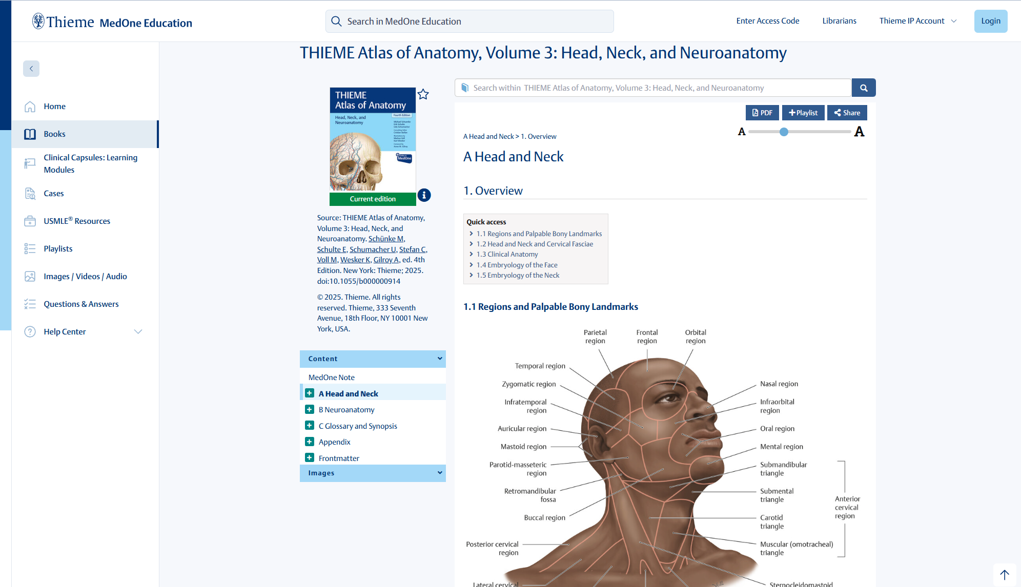
THIEME Atlas of Anatomy, Vol. 3
Head, Neck, and Neuroanatomy, 4th Edition
Schuenke | Schulte | Schumacher | Stefan
Publication Date: 2025 | Edition: 4 | 628pp, 1,801 illustrations
Print ISBN: 9781684205943 | E-Book ISBN: 9781684205967
Exceptional atlas combines highly detailed illustrations with relevant applied and clinical anatomy
This three-in-one atlas combines exquisite illustrations, brief descriptive text/tables, and clinical applications, making it an invaluable instructor- and student-friendly resource for lectures and exam prep. Head and neck sections encompass the bones, ligaments, joints, muscles, lymphatic system, organs, related neurovascular structures, and topographical and sectional anatomy. The neuroanatomy section covers the histology of nerve and glial cells and autonomic nervous system, then delineates different areas of the brain and spinal cord, followed by sectional anatomy and functional systems. The final section features a glossary and CNS synopses.
Key Features:
- More than 1,800 extraordinarily accurate and beautiful illustrations by Markus Voll and Karl Wesker enhance understanding of anatomy
- A significant number of images have been revised to reflect gender and ethnic diversity
- Superb topographical illustrations support dissection in the lab
- Two-page spreads provide a teaching and learning tool for a wide range of single anatomic concepts
This visually stunning atlas is an essential companion for medical students or residents interested in pursuing head and neck subspecialties or furthering their knowledge of neuroanatomy. Dental and physical therapy students, as well as physicians and physical therapists seeking an image-rich, clinical practice resource will also benefit from consulting this remarkable atlas.
What MedOne Education can do
For the student
- NInstant access all required medical textbooks
- NIdeal for quick reference or in-depth review
- NEasily accessible – even offline – the perfect study companion
- NCovers the clinical and scientific topics your medical students need to succeed
For the instructor
- NRequire titles with no cost to students
- NEasily download images for lectures
- NCreate custom playlists for every course
- NAdd notes for future reference
For the institution
- NInvite students to learn anytime or place
- NImprove efficient teaching
- NAppeal to prospective students and faculty
- NSave time through high user-friendliness
Already have access? If your institution already has access to MedOne Education, please visit: medone-education.thieme.com
